
Wireless Microimplants for Deep Tissue Disease Monitoring and Treatment
Principal Investigator: Professor O’Sullivan
AWaRE REU Researcher: Anuj Gajjar, Northeastern University
Other Contributors: Alicia Wei
Project Summary:
Despite the explosive growth in the development of wearable and implantable sensors for monitoring health and personal wellness, there are currently no viable sensor technologies that can sense targets deep within the human body. Current sensors are limited to sensing cutaneous or shallow subcutaneous tissue volumes, have limited functionality, or are simply too large and obtrusive. This prevents their use in some of the most impactful areas of medicine including monitoring solid tumors, simultaneous deep brain sensing and stimulation, and monitoring diseases of the internal organs. The long-term goal of this project is to develop an extensible microimplant platform that can ultimately be placed anywhere in the human body and provide sensitivity to multiple biomolecular targets continuously and in real-time. This project entails sensor design, modeling (mechanical and functional), and prototyping of wireless microimplants using advanced manufacturing processes.
Finding:
Breast tissue markers are tiny radiologically-visible grain-sized implants which are routinely implanted into suspicious breast lesions during biopsy procedures. These “breast clips” enable clinicians to radiologically locate breast lesions and group diseased areas for more precise monitoring. We are developing similarly-sized “smart” breast clips that can provide real-time information of a tumor to optimize treatment and extend survival. These smart clips consist of hyperspectral optical sources, photodetectors, and wireless power receiver and communication circuitry. These hyperspectral sources detect changes in concentrations and chemical states of three biomarkers: hemoglobin (oxygenated and deoxygenated), water (protein-bound or free), and lipids (saturated or unsaturated), which are directly related to tumor composition, metabolism, and vascularity. Such biomarkers can also predict a pathologic complete response (pCR) to chemotherapy.
The smart breast clip performs this molecular sensing of tumor composition and hemodynamics via two microchip vertical-cavity surface-emitting lasers (VCSELs) covering the optical bandwidth (visible and near-infrared light) used to measure the above biomarkers. The implant also includes an analog front end to control the VCSELs and photodetectors, a receiver coil and impedance matching and rectification circuitry for wireless power transfer, as well as load-modulation of the RF power field for wireless communication. All sensor components and circuitry are integrated into a single printed circuit board sized to standard syringe needle sizes for in vivo implantation.
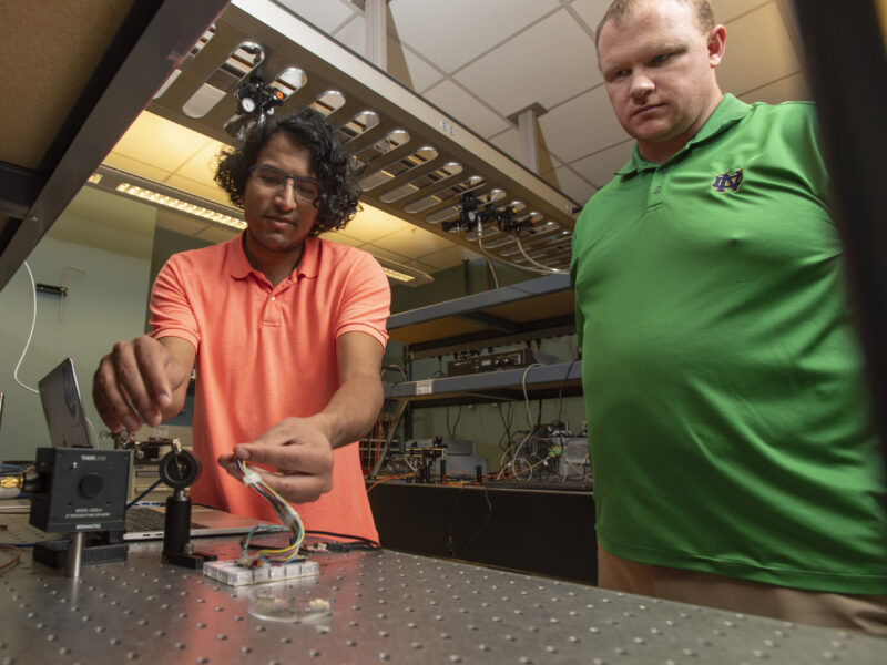
To enable the final development aim of preclinical evaluation of the device, the accuracy, precision, dynamic range, stability, and tissue depth range must be well characterized. This characterization involves simulating the optical performance of the sensor, profiling the power performance of the VCSELs, profiling the response of the sensor’s photodetectors, and testing performance in an optical and electromagnetic phantom. In this poster, we describe our work to enable repeatable characterization of smart breast clips.
The first goal was to perform Monte-Carlo-based tissue optical simulations of the device to obtain metrics about the overall expected optical performance of the breast clip. The second goal of this work was to automate a characterization procedure for the breast clip, which involved reading measurements from various external detectors that characterize laser and photodetector performance, as well as controlling the analog front end within the sensor. Using a combination of Python and C++ programming, a modular software system was built to achieve both goals, and thus provide progress toward preclinical evaluation of the breast clip.